Fungal biomass estimation based on ergosterol extraction
Raimbault A.1; Erdozain M.1; de-Miguel S.1, 2
1 Forest Science and Technology Centre of Catalonia
2 Department of Agricultural and Forest Sciences and Engineering, University of Lleida, Lleida, Spain
Last updated: 25/09/2026
Recommended citation:
Raimbault A.; Erdozain M.; de-Miguel S. 2026. Fungal biomass estimation based on ergosterol extraction. In: Pere Casals, Davide Ascoli, Lena Vilà-Vilardell, Imma Oliveras, Madoka Krick (eds.) 2026. Methods handbook for characterizing fire use behavior, assessing fire effects, and monitoring soil and vegetation responses at the stand level. FIRE-ADAPT project (number 101086416), deliverable D1.1
Indicator
Ecosystem responses: Soil responses
Brief introduction
The structure of soil microbial communities plays an essential role in the decomposition and utilization of soil organic matter by influencing metabolic performance (Bailey et al., 2002; Allison et al., 2013). Within the microbial community, fungi are thought to be more efficient than bacteria in stabilizing and storing carbon in soils (Malik et al., 2016). However, it has been demonstrated that wildfires affect microbial biomass, including fungal biomass (Holden and Treseder, 2013). Indeed, the combustion of aboveground vegetation and soil organic matter leads to the loss of large amounts of organic carbon (Amiro et al., 2001; Van Der Werf et al., 2010), leaving behind a more recalcitrant organic carbon that is therefore more difficult for microbes to decompose (Holden and Treseder, 2013). Estimating fungal biomass is thus an effective way to assess the health and productivity of soil ecosystems and would help predict the consequences of environmental changes and management practices on carbon dynamics (Martinovic et al., 2024).
Several methods exist for measuring microbial biomass in forest soils, each with its own advantages and limitations (Martinovic et al., 2024). The first is Fumigation-Extraction, which enables direct quantification of total microbial biomass and is widely recognized in the scientific community. However, it does not allow bacteria and fungi to be distinguished and tends to underestimate actual biomass (Brookes et al., 1985; Xu et al., 2013; Martinovic et al., 2024). The second approach, PLFA (phospholipid fatty acid analysis), allows the biomass of specific microbial groups to be estimated with high precision, although it requires specialized equipment (Frostegård et al., 1991; Joergensen, 2022; Martinovic et al., 2024). The third approach, qPCR (quantitative polymerase chain reaction) is a molecular approach that amplifies and quantifies DNA from environmental samples. While this method offers high sensitivity, it requires prior knowledge of the target sequences and remains susceptible to handling errors, particularly in the presence of contaminants (Smith and Osborn, 2009; Martinovic et al., 2024). Finally, the last approach consists in the extraction of ergosterol, a component of the cell membrane of mycorrhizal fungi, which is frequently used as a biomarker to estimate fungal biomass (Wilkes, 2023). Several extraction methods exist, involving the use of potentially hazardous chemicals, with varying exposure times depending on the protocol employed (Wilkes, 2023).
This protocol is divided into two distinct steps. The first step concerns soil sampling and is based on the protocol established by Martinovic et al. (2024). The second step, dedicated to ergosterol extraction, draws on the work of Wilkes (2023), in which different ergosterol extraction methods were compared. The present protocol is based on the conclusions of this study and adopts the method that considerably reduces chemical hazard exposure for the user. It therefore describes ergosterol extraction using chloroform.
Method specifications
Method type
Soft
Hard
Method timing
Before
fire
During
fire
After fire
(short-term)
After fire
(mid-term)
After fire
(long-term)
Field skills
Low
Medium
Hard
Lab skills
Low
Medium
Hard
Material cost
Low
Medium
Hard
Time cost
Low
Medium
Hard
Calculation expertise
Low
Medium
Hard
Equipment
- Auger
- Knife (or other tool for extracting soil from the corer)
- Balance
- Notebook
- Sieve for soil with 5-mm mesh size
- Plastic 50-ml plastic tubes (or alternatively plastic ziploc bags with 50 ml volume)
- Latex glove
- Ruler
- Permanent gloves
- Ventilated drying oven
- Analytical balance
- Heated ultrasonic bath
- Centrifuge with rotor accommodating 50 ml tubes
- Water bath
- Fume hood
- Micropipettes
- 50 ml centrifuge tubes
- Glass Pasteur pipettes
- Volumetric glassware, spatulas, tube racks
- Chloroform (analytical grade)
- Methanol (analytical grade and HPLC grade)
- Laboratory coat, safety goggles, laminate gloves (nitrile is permeable to chloroform)
Experimental design
The main objective is to ensure sufficient coverage to represent the entire burned area and capture its environmental variability. If the burned area is large (>1 ha) and the resources allow it, several plots within the burned area should be selected (at least 3), in ecologically contrasting sites (e.g. in terms of pH, soil moisture or vegetation type) to better reflect broader environmental gradients. When comparing burned to control plots, however, it is important to ensure that the selected plots between these areas are as comparable as possible in terms of characteristics such as geology, topography, vegetation (pre-burned) and climate.
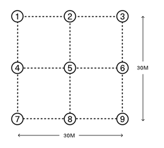
Collect a minimum of 4 sub-samples per plot, spaced at least 2 m apart, and pool them into a single plastic bag (Štursová et al., 2016; Martinovic et al., 2024). This design captures the spatial heterogeneity of the plot while remaining compatible with the available resources (time, personnel and field accessibility). Record the GPS coordinates of each sampling area. The spatial configuration of the subsamples within each plot can follow established protocols such as those developed by SPUN (900 m²) (SPUN, 2026) and HoliSoil (16 m²) (Baldrian et al., 2024), which recommend very different sampling surface areas (Figure 1).
Methodology
Soil Sampling
- Prior to sampling, it is necessary to prepare sterile 50 ml tubes by labelling them with a unique plot code, written both on the cap and on the tube. The empty mass of each tube must also be recorded in advance, with a resolution of 0.01 g if possible.
- The surface litter layer should be removed prior to sampling, and soil should be collected from the top 10 centimetres with an auger, where approximately 95% of belowground biomass is concentrated (Šnajdr et al., 2008).
- It is recommended to wear latex gloves throughout the handling of soil samples. During the extraction of soil cores, stones and roots should be removed using a wooden rod. To eliminate remaining debris, the samples should then be sieved. Any residual fine debris can be removed using tweezers.
- The composite sample is prepared by thoroughly mixing all soil sub-samples in a plastic bag. The 50 ml tube is then filled up to 40 ml and the wet mass of the whole (tube + soil) is immediately recorded.
- While awaiting biomass analyses, tubes can be stored in a freezer at a temperature of -20°C.
Ergosterol extraction
- Prior to extraction, samples should be air-dried at 25 °C for a minimum of 48 hours. Samples are considered sufficiently dry when three consecutive weight measurements, taken at least 6 hours apart, yield stable and consistent values.
- This protocol is a modified version of the method proposed by Salazar Alekseyeva et al. (2021). Weigh 300 mg of sample into 50 ml centrifuge tubes.
- Add 3 ml of a chloroform/methanol mixture1 (2:1) to each sample, then sonicate (ultrasonic agitation) for 30 minutes at 50 °C in a tightly closed tube.
- Allow the samples to cool to room temperature, then incubate at room temperature for 18 hours.
- Perform a second sonication at 50 °C for 20 minutes, followed by centrifugation at 1000 g for 1 minute.
- Collect the supernatant (the upper liquid phase) and transfer it to a clean tube.
- Heat the tube continuously in a water bath at 40 °C until complete evaporation of the methanol and chloroform.
- Add 1 ml of HPLC-grade methanol to each tube and incubate at 40 °C for 15 minutes.
- Filter the solution through 0.2 µm nylon membrane syringe filters (Chromatography Direct) into amber glass HPLC vials until analysis2.
1 All chemicals and reagents can be purchased from Thermo Fisher Scientific.
2 It is strongly recommended to continuously monitor volatile organic compound (VOC) exposure in the vicinity of the user using an appropriate detector (e.g., ToxiRAE Pro PID, Honeywell).
Calculations
Following HPLC analysis according to the protocols of Mille-Lindblom et al. (2004) and Wilkes et al. (2021), which allow quantification of ergosterol concentration in the samples, fungal biomass can be estimated using the following equation (Montgomery et al., 2000) 3 :
\mathrm{FB}(\mu\mathrm{g/g})= \mathrm{Ergosterol}(\mu\mathrm{g/g})\times f\times R_f
3 This conversion factor implies a constant ratio between ergosterol content and fungal mycelial mass, which is unlikely to hold across samples given interspecific variability and differences in environmental conditions (Montgomery et al., 2000; Wilkes, 2023). The equation is also inappropriate for arbuscular mycorrhizal fungi, which do not typically contain ergosterol (Olsson et al., 2003).
Relation with other methods
NA
Illustrations

Data templates
NA
Further information
Allison, S. D., Lu, Y., Weihe, C., Goulden, M. L., Martiny, A. C., Treseder, K. K., & Martiny, J. B. (2013). Microbial abundance and composition influence litter decomposition response to environmental change. Ecology, 94(3), 714-725.
Amiro, B. D., Stocks, B. J., Alexander, M. E., Flannigan, M. D., & Wotton, B. M. (2001). Fire, climate change, carbon and fuel management in the Canadian boreal forest. International Journal of Wildland Fire, 10(4), 405-413.
Bailey, V. L., Smith, J. L., & Bolton Jr, H. J. S. B. (2002). Fungal-to-bacterial ratios in soils investigated for enhanced C sequestration. Soil Biology and Biochemistry, 34(7), 997-1007.
Baldrian, P., Martinovic, T., Fort, T., Adamczyk, B., Weikl, F., Pritsch, K., Brabcová, V., 2024. Harmonised methodologies for analysis of soil biodiversity, functional redundancy & community resilience in forest soils. HoliSoils project. Deliverable 1.4. file:///D:/40879755S/Desktop/FireAdapt/Attachment_0.pdf
Brookes, P.C., Landman, A., Pruden, G., Jenkinson, D.S., 1985. Chloroform fumigation and the release of soil nitrogen: A rapid direct extraction method to measure microbial biomass nitrogen in soil. Soil Biology and Biochemistry 17, 837–842.
Frostegård, Å., Bååth, E., Tunlid, A., 1993. Shifts in the structure of soil microbial communities in limed forests as revealed by phospholipid fatty acid analysis. Soil Biology and Biochemistry 25, 723–730.
Holden, S. R., & Treseder, K. K. (2013). A meta-analysis of soil microbial biomass responses to forest disturbances. Frontiers in microbiology, 4, 163.
Joergensen, R.G., 2022. Phospholipid fatty acids in soil—drawbacks and future prospects. Biology and Fertility of Soils 58, 1–6.
Malik, A. A., Chowdhury, S., Schlager, V., Oliver, A., Puissant, J., Vazquez, P. G., … & Gleixner, G. (2016). Soil fungal: bacterial ratios are linked to altered carbon cycling. Frontiers in microbiology, 7, 1247.
Martinovic, T., Baldrian, P., Santonja, M., Izquierdo, L. P., & Confidential, C. O. (2024). D1. 3-Harmonised methodologies for quantification of biomass of soil organisms.
Mille-Lindblom, C., Von Wachenfeldt, E., & Tranvik, L. J. (2004). Ergosterol as a measure of living fungal biomass: persistence in environmental samples after fungal death. Journal of microbiological methods, 59(2), 253-262.
Montgomery, H. J., Monreal, C. M., Young, J. C., & Seifert, K. A. (2000). Determinination of soil fungal biomass from soil ergosterol analyses. Soil Biology and Biochemistry, 32(8-9), 1207-1217.
Olsson, P. A., Larsson, L., Bago, B., Wallander, H., & Van Aarle, I. M. (2003). Ergosterol and fatty acids for biomass estimation of mycorrhizal fungi. New Phytologist, 7-10.
Salazar Alekseyeva, K.; Mähnert, B.; Berthiller, F.; Breyer, E.; Herndl, G.J.; Baltar, F. Adapting an Ergosterol Extraction Method with Marine Yeasts for the Quantification of Oceanic Fungal Biomass. J. Fungi 2021, 7, 690. https://doi.org/10.3390/jof7090690
Smith, C.J., Osborn, A.M., 2009. Advantages and limitations of quantitative PCR (Q-PCR)-based approaches in microbial ecology. FEMS Microbiology Ecology 67, 6–20.
Šnajdr, J., Valášková, V., Merhautová, V.r., Herinková, J., Cajthaml, T., Baldrian, P., 2008. Spatial variability of enzyme activities and microbial biomass in the upper layers of Quercus petraea forest soil. Soil Biology and Biochemistry 40, 2068-2075.
SPUN, 2026. Sampling Protocol, Sampling of soil for molecular analysis of AMF and EMF DNA sequences modified from the Silva Nova/SoilBon protocol. In.
Štursová, M., Bárta, J., Šantrůčková, H., Baldrian, P., 2016. Small-scale spatial heterogeneity of ecosystem properties, microbial community composition and microbial activities in a temperate mountain forest soil. FEMS Microbiology Ecology 92, fiw185.
Van der Werf, G. R., Randerson, J. T., Giglio, L., Collatz, G. J., Mu, M., Kasibhatla, P. S., … & van Leeuwen, T. T. (2010). Global fire emissions and the contribution of deforestation, savanna, forest, agricultural, and peat fires (1997–2009). Atmospheric chemistry and physics, 10(23), 11707-11735.
Wilkes, T. I., Warner, D. J., Edmonds-Brown, V., Davies, K. G., & Denholm, I. (2021). Zero tillage systems conserve arbuscular mycorrhizal fungi, enhancing soil glomalin and water stable aggregates with implications for soil stability. Soil Systems, 5(1), 4.
Wilkes, T. I. (2023). Ergosterol extraction: a comparison of methodologies. Access microbiology, 5(4), 000490-v4.
Xu, X., Thornton, P.E., Post, W.M., 2013. A global analysis of soil microbial biomass carbon, nitrogen and phosphorus in terrestrial ecosystems. Global Ecology and Biogeography 22, 737– 749.